Fundus Autofluorescence
What is Fundus Autofluorescence?
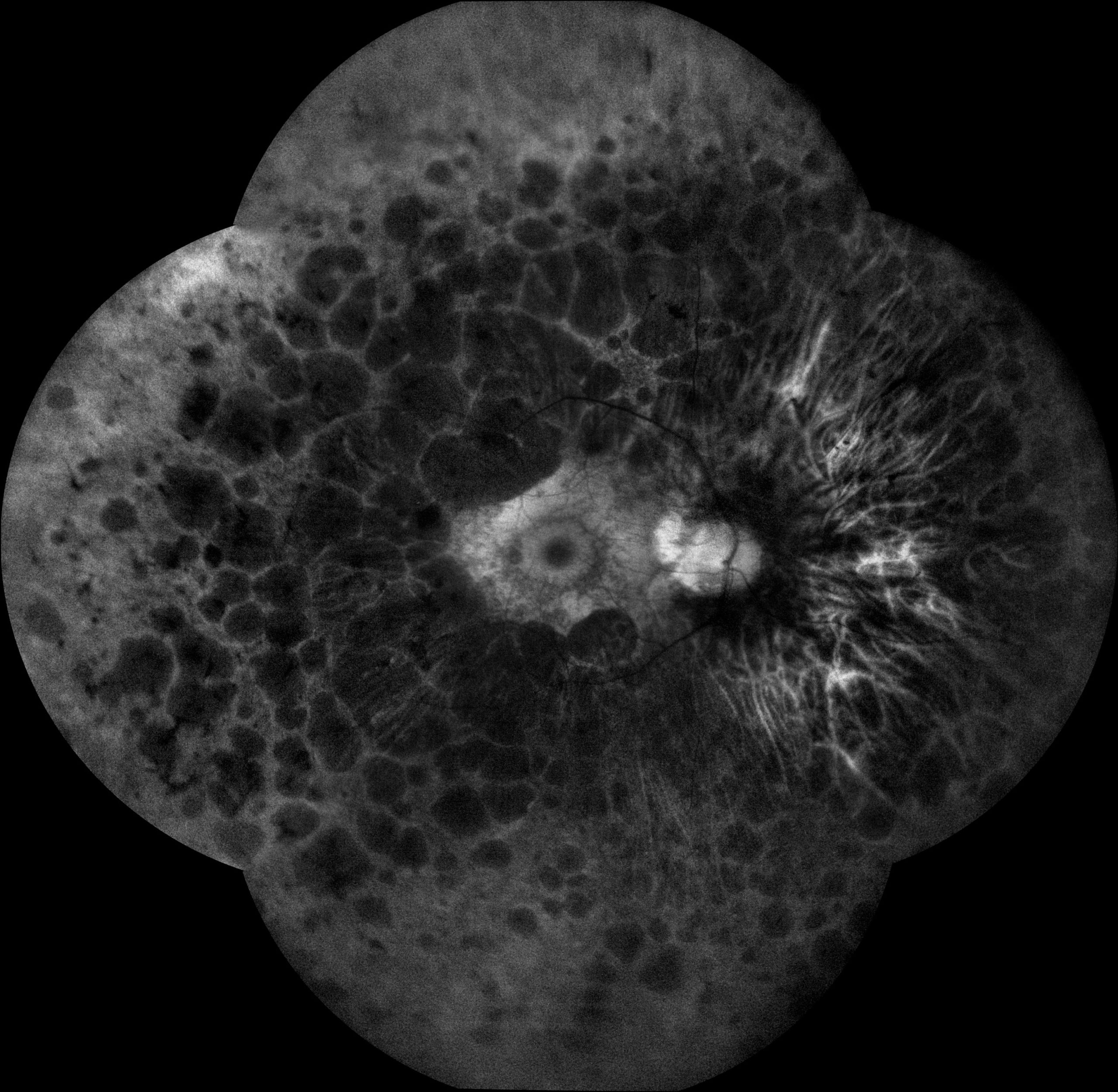
Fundus autofluorescence (FAF) imaging is a non-invasive imaging technique used to visualize the distribution and intensity of natural fluorescence emitted by retinal pigment epithelial (RPE) cells in the back of the eye. It provides valuable information about the health and function of the RPE layer, which plays a crucial role in maintaining the structural integrity and function of the retina.
Fundamentals of Fundus Autofluorescence
Here’s how fundus autofluorescence works and its significance in clinical practice:
- Principle of autofluorescence: The RPE cells contain lipofuscin, a naturally occurring pigment that accumulates with age and is a byproduct of the visual cycle. When excited by blue or near-ultraviolet light, lipofuscin emits fluorescence in the green wavelength range. Fundus autofluorescence imaging captures this emitted fluorescence to create detailed images of the distribution and intensity of lipofuscin within the RPE layer.
- Image capture: Fundus autofluorescence imaging is typically performed using a specialized fundus camera equipped with filters to detect the emitted fluorescence from the RPE. The patient’s pupils are dilated, and the camera captures images of the back of the eye in a darkened room.
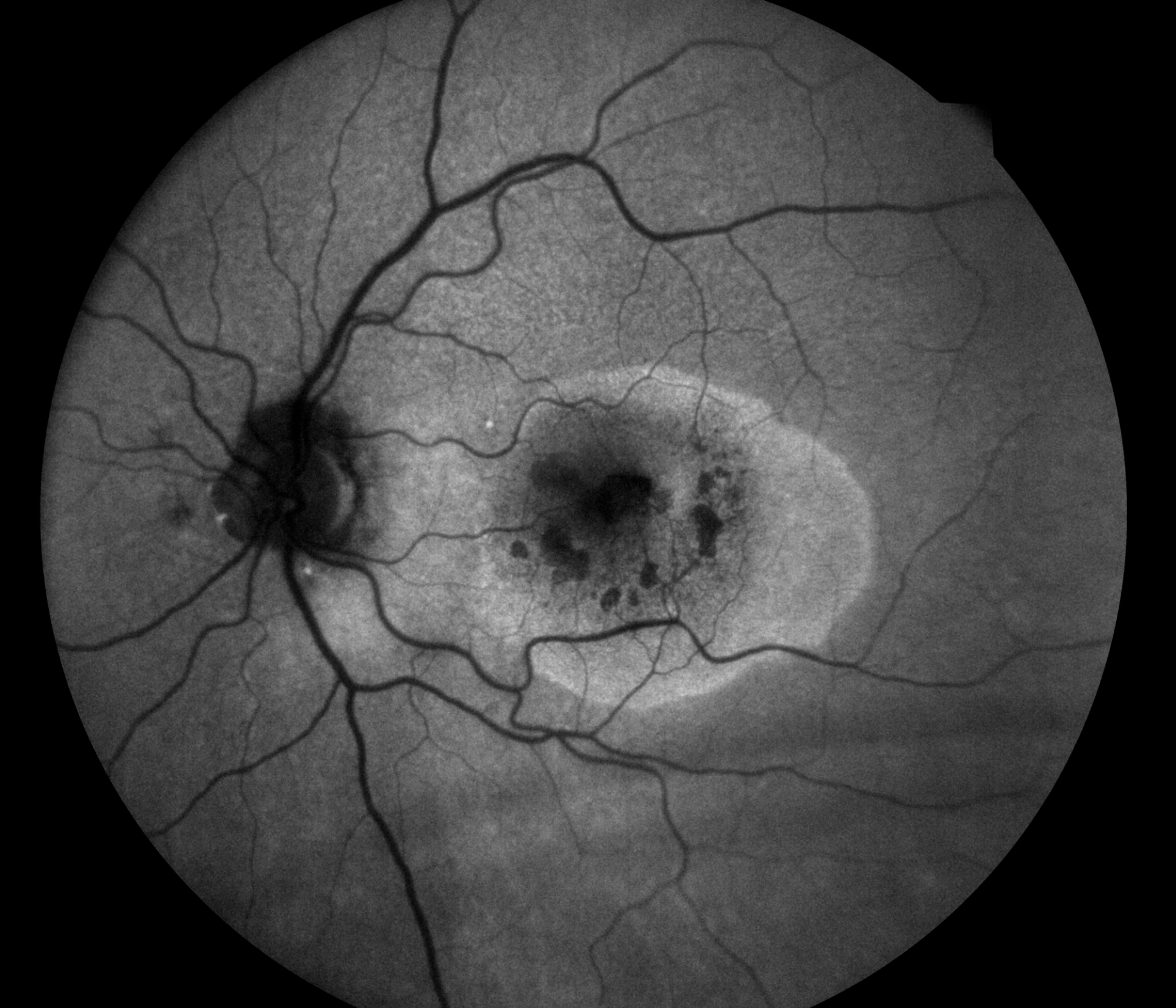
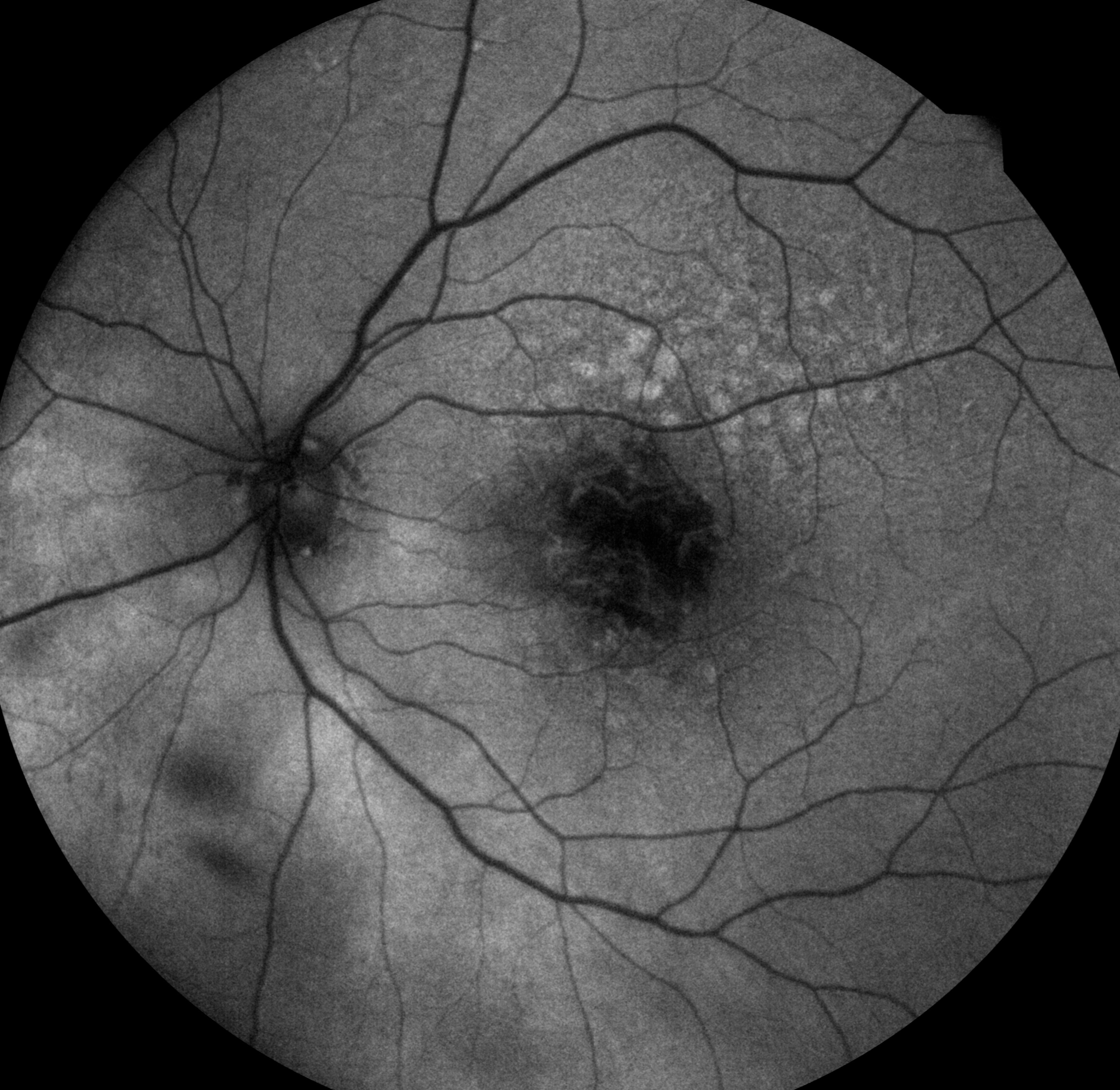
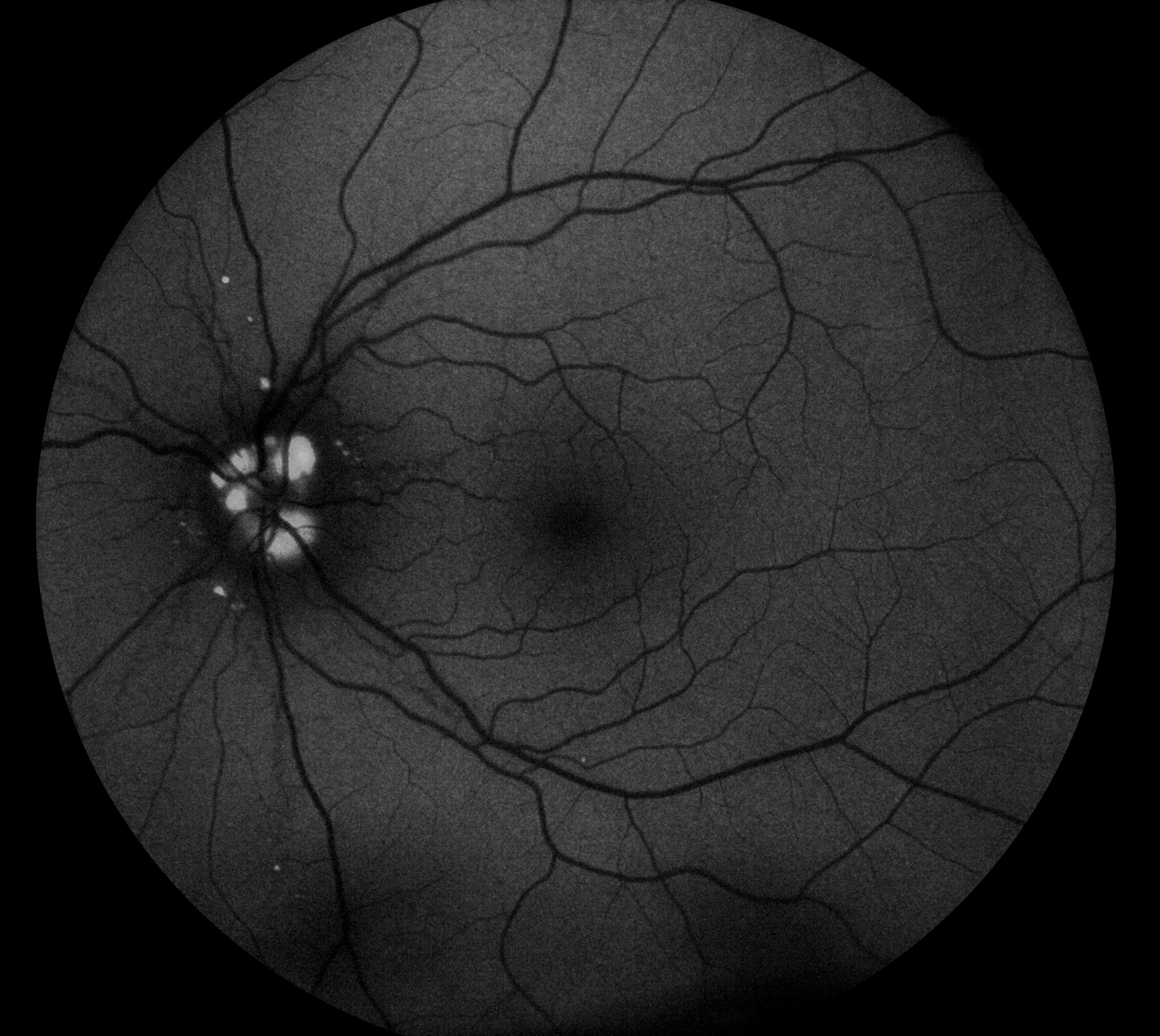
- Analysis: The captured images are then analyzed by retinal specialists. Areas of increased autofluorescence typically indicate regions of higher lipofuscin accumulation within the RPE layer, while areas of decreased autofluorescence may indicate loss or dysfunction of RPE cells. Changes in autofluorescence patterns can provide valuable diagnostic information about various retinal diseases and conditions.
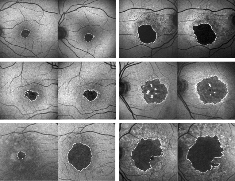
- Clinical applications: Fundus autofluorescence imaging is used in the diagnosis, monitoring, and management of a variety of retinal disorders. It can help identify and assess the extent of geographic atrophy in conditions such as age-related macular degeneration (AMD), where there is loss of RPE cells and photoreceptors. In diseases such as central serous chorioretinopathy (CSCR), fundus autofluorescence can reveal areas of RPE dysfunction and focal leakage. Autofluorescence patterns can also aid in the diagnosis and monitoring of inherited retinal dystrophies, such as retinitis pigmentosa and Stargardt disease.
- Treatment planning: Fundus autofluorescence imaging can provide valuable insights for treatment planning in retinal diseases. By identifying areas of RPE dysfunction or atrophy, clinicians can better target therapeutic interventions such as anti-vascular endothelial growth factor (anti-VEGF) injections, photodynamic therapy, or gene therapy. It can also help monitor treatment response and disease progression over time.