Scleral Buckle Surgery
What is Scleral Buckle Surgery
Scleral buckle surgery is a procedure used to repair certain types of retinal
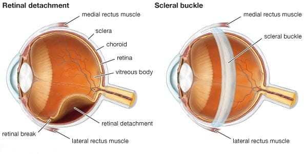
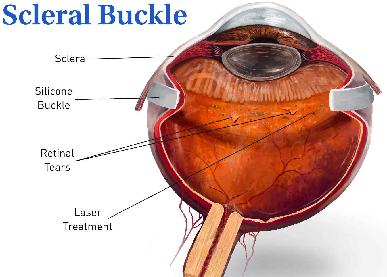
detachments. It involves the placement of a silicone band (the scleral buckle) around the outer wall of the eye (the sclera) to support the detached retina and help it reattach to the back of the eye. This procedure is often performed in a hospital or surgical center under local or general anesthesia.
Process for Scleral Buckle Surgery
Here’s an overview of the steps involved in scleral buckle surgery:
- Preparation: Before the surgery, your eye will be numbed with local anesthesia, and you may also receive a sedative to help you relax or be put fully to sleep under general anesthesia. Your pupils will be dilated to provide better visualization to the inside of the eye.
- Incisions: Dr. Haak will make one or more small incisions in the conjunctiva (the thin, transparent membrane covering the white part of the eye) to expose the sclera.
- Placement of scleral buckle: A silicone band or sponge is then placed around the circumference of the eye, just behind the muscles that control eye movement. The buckle is secured in place with sutures.
- Draining fluid: If there is fluid or blood under the retina, Dr. Haak may drain it using a small needle or drainage tube inserted through the sclera.
- Indentation: The scleral buckle creates an indentation or indentation in the wall of the eye, which helps to push the wall of the eye inward and reduce the force pulling on the retina. This indentation also helps to close any retinal tears or breaks and allows the retina to reattach to the back of the eye.
- Closing incisions: Once the scleral buckle is in place and any necessary procedures are completed, the incisions in the conjunctiva are closed with sutures.
After scleral buckle surgery, you may experience some discomfort, swelling, or redness in the eye, which can usually be managed with over-the-counter pain medication such as Tylenol and prescription eye drops. Your vision may also be blurry or distorted initially, but it typically improve as the eye heals and the retina reattaches.
It is likely that you may require a prescription change for glasses after the
procedure.
It’s important to follow the postoperative care instructions, including attending follow-up appointments to monitor the healing process and ensure that the retinal detachment is resolving properly. In some cases, additional procedures or treatments may be needed to achieve the best possible outcome. Flying or gain of altitude may be limited for a certain period if intraocular gas/air is utilized.


