Fundus Photography
What is Fundus Photography?
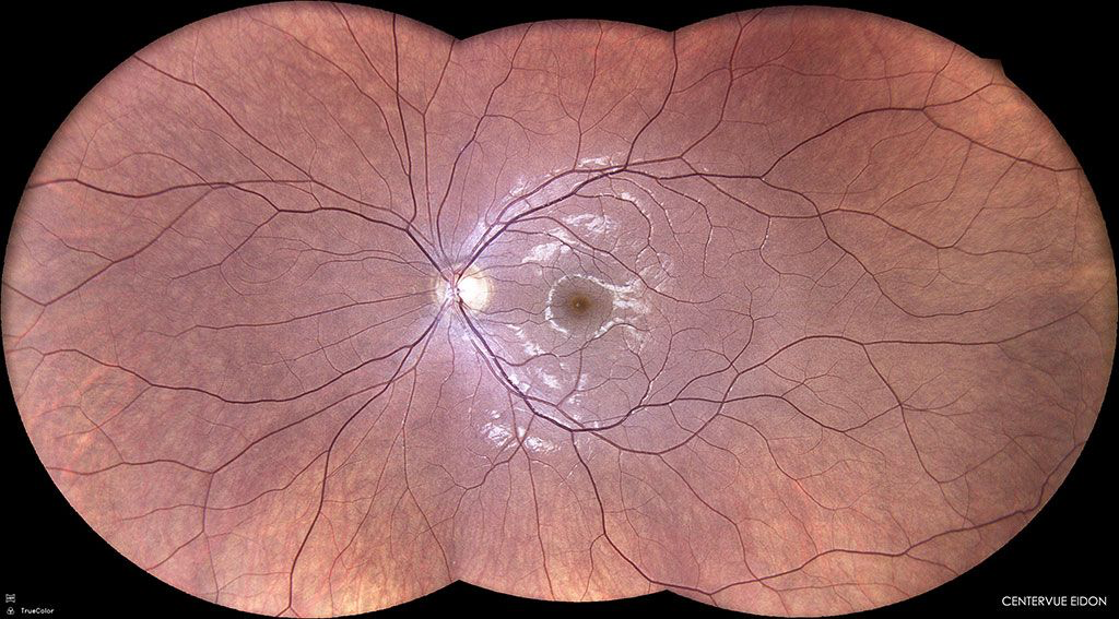
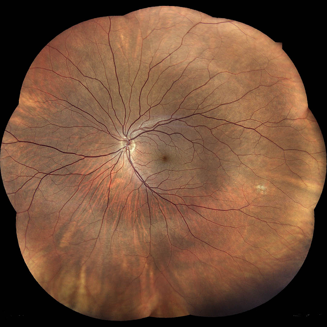
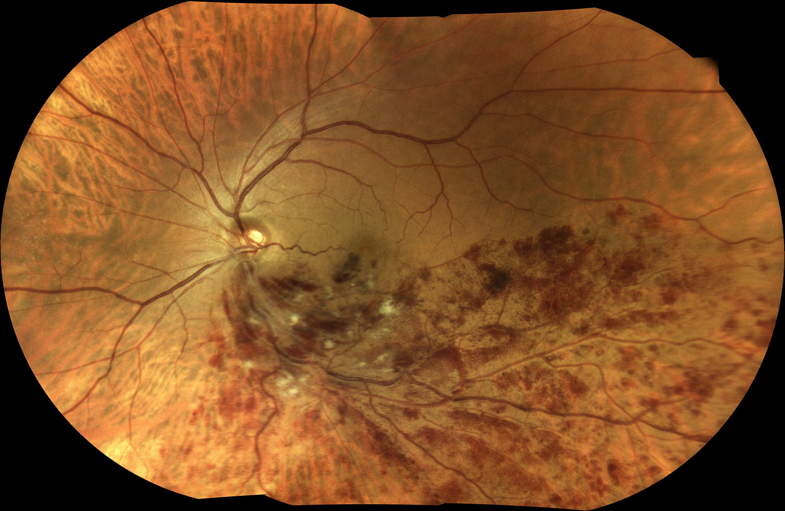
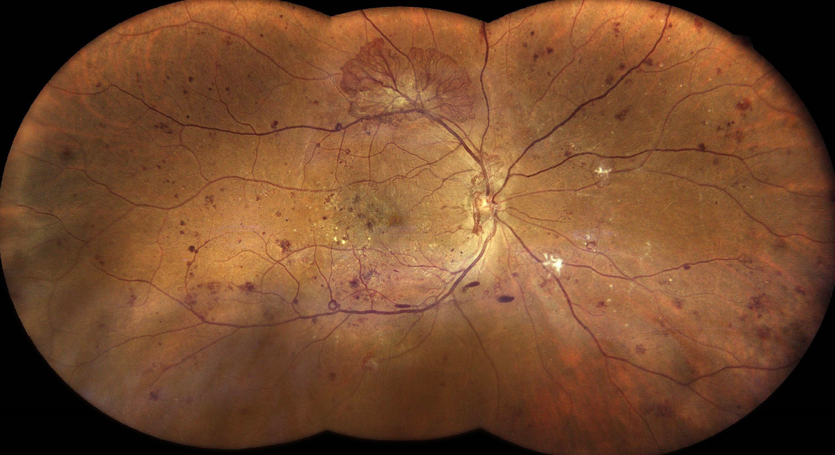
Fundus photography is a specialized imaging technique used to capture detailed photographs of the back of the eye, including the retina, optic nerve, and macula. It provides high-resolution images of the fundus, which is the interior surface of the eye opposite the lens, allowing retina specialists to assess the health and condition of these critical structures.
Fundamentals of Fundus Photography
Here’s how fundus photography works and its significance in clinical practice:
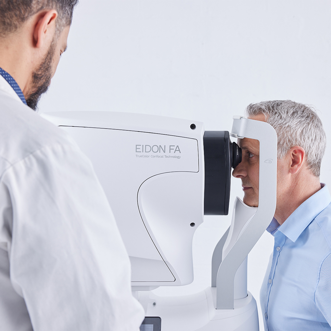
- Equipment: Fundus photography typically involves the use of a fundus camera, which is a specialized camera equipped with filters and lenses designed to capture images of the retina and surrounding structures.
- Image capture: The patient is positioned comfortably in front of the fundus camera, and the photographer adjusts the settings to optimize image quality. The camera emits a flash of light, which illuminates the interior of the eye, allowing the camera to capture detailed photographs of the fundus.
- Image analysis: The captured images are then reviewed and analyzed by the retina specialist. The images are examined for signs of retinal pathology, such as diabetic retinopathy, age-related macular degeneration (AMD), retinal vein occlusions, hypertensive retinopathy, retinal detachments, and other conditions. Fundus photography provides valuable information about the appearance, location, and severity of retinal abnormalities, guiding diagnosis, treatment planning, and monitoring of disease progression.
- Documentation and monitoring: Fundus photography serves as a valuable tool for documenting baseline retinal findings and monitoring changes over time. Serial fundus photographs allow clinicians to track the progression of retinal diseases, evaluate the effectiveness of treatment interventions, and make informed decisions about patient management.
- Patient education: Fundus photographs can also be used to educate patients about their eye health and the impact of retinal conditions on their vision. By visually illustrating the presence and severity of retinal pathology, patients gain a better understanding of their condition and the importance of adhering to treatment recommendations and lifestyle modifications.
Overall, fundus photography is an essential diagnostic tool in ophthalmology, providing detailed images of the retina and optic nerve that aid in the early detection, diagnosis, and management of a wide range of ocular diseases. Its non-invasive nature, high image quality, and ability to track changes over time make it an invaluable asset in evaluation, management, and follow up of retinal conditions.