Vitrectomy Surgery
What is Vitrectomy Surgery?
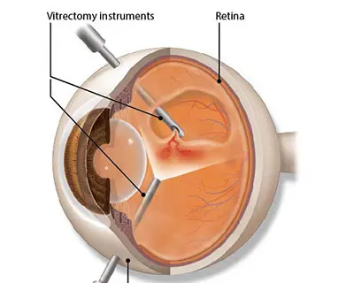
Vitrectomy surgery is a procedure performed to remove some or all of the vitreous gel from the middle of the eye. The vitreous gel is a clear, gel-like substance that fills the space between the lens and the retina. Vitrectomy surgery is commonly used to treat a variety of eye conditions affecting the vitreous, retina, and other structures within the eye.
Process for Vitrectomy Surgery
Here’s an overview of the procedure:
- Preparation: Before the surgery, you will typically undergo a thorough eye examination and various diagnostic tests to evaluate the condition of your eye and determine if vitrectomy surgery is the appropriate treatment.
- Anesthesia: Vitrectomy surgery is usually performed under local or general anesthesia, depending on the patient’s preference and the complexity of the procedure.
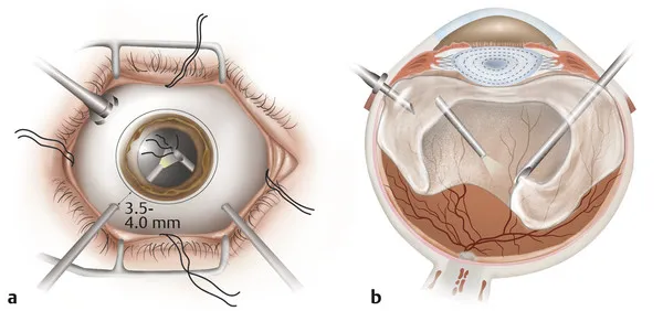
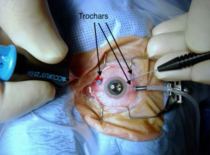
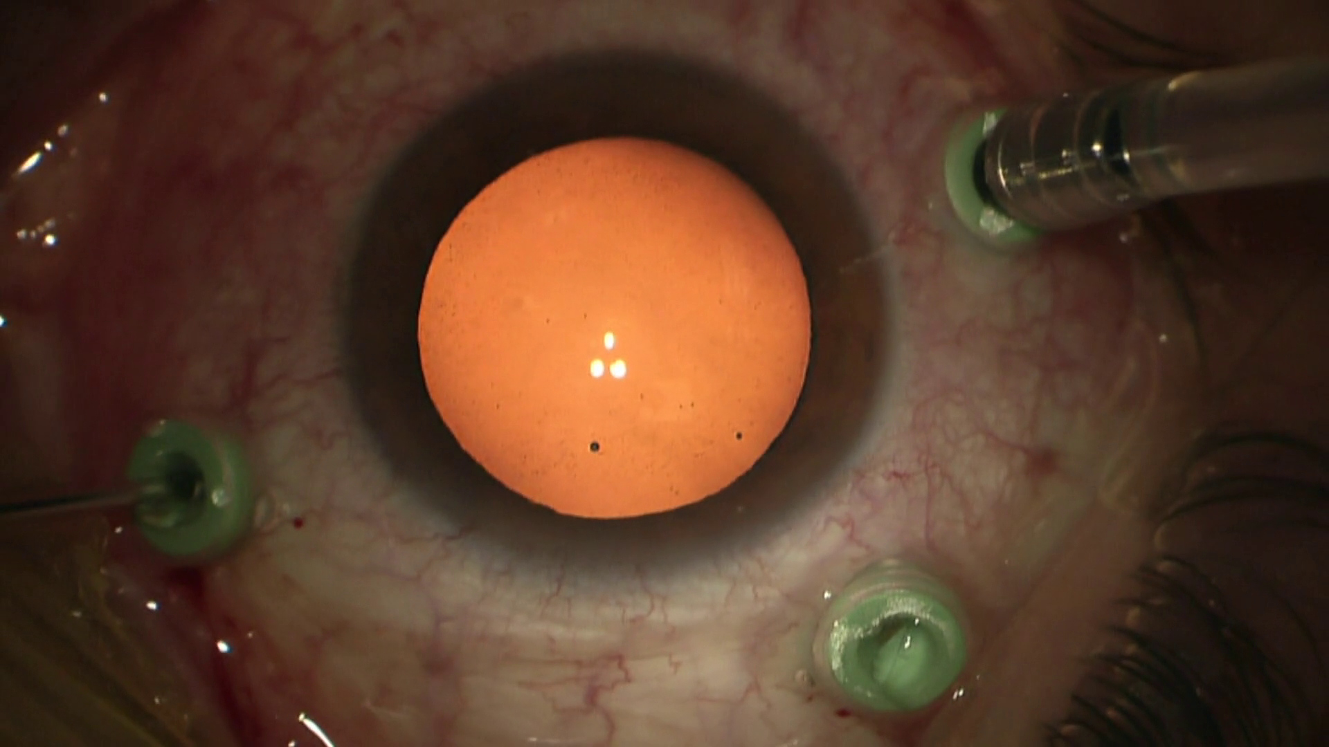
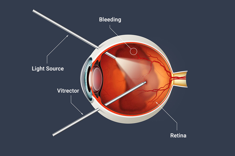
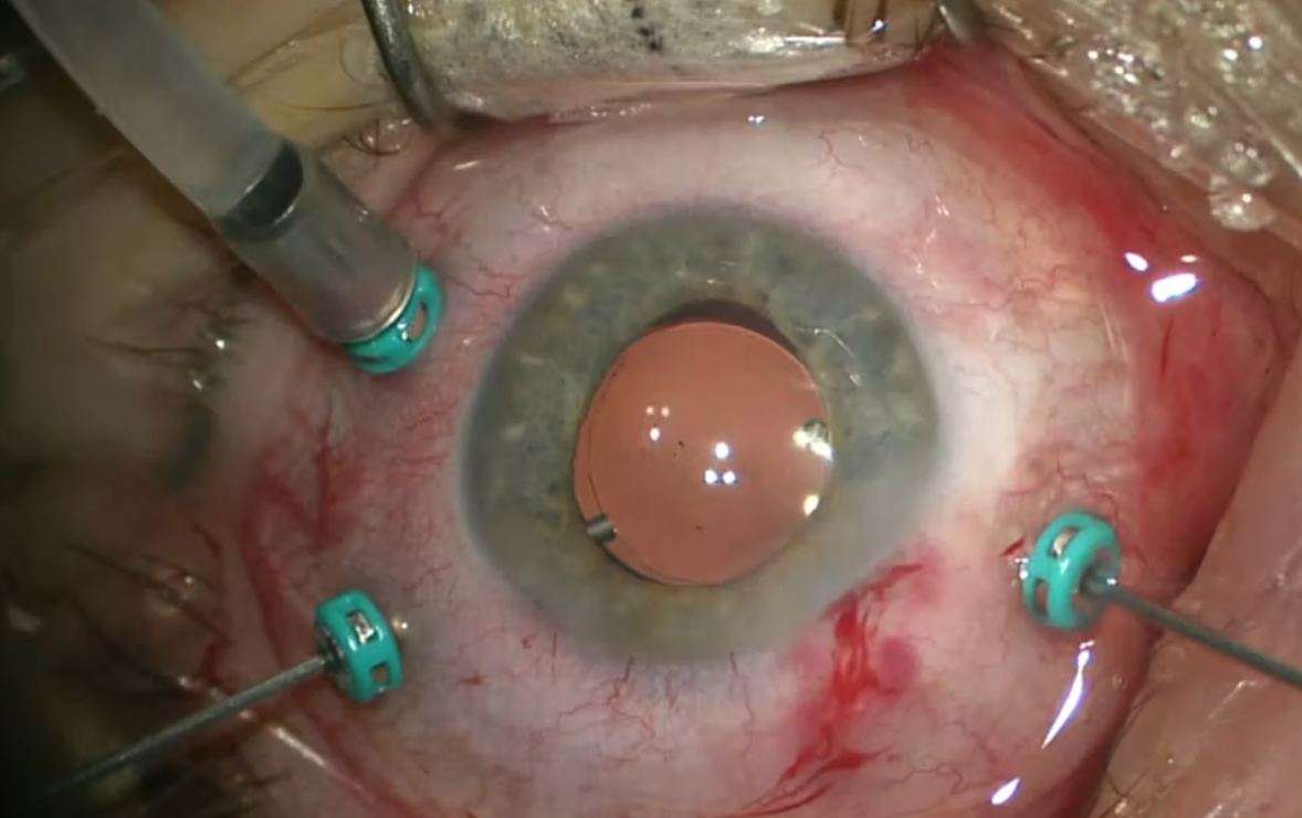
- Incisions: Dr. Haak will make small incisions in the sclera (the white part of the eye) to access the vitreous cavity. These incisions are typically very small and may be sutured or left to heal on their own.
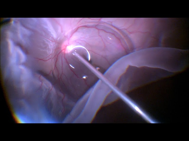
- Vitrectomy: Using specialized instruments, including a vitrectomy probe, light source, and infusion system, Dr. Haak will remove the vitreous gel from the eye. This may involve removing only a portion of the vitreous gel or completely removing it, depending on the specific needs of the patient and the underlying condition being treated.
- Treatment of underlying conditions: In addition to vitreous removal, Dr. Haak may perform other procedures during the vitrectomy to address underlying eye conditions. For example, they may repair retinal tears or detachments, remove scar tissue, or treat abnormal blood vessel growth.
- Closure: Once the vitrectomy and any additional procedures are completed, the incisions may be closed with sutures or allowed to heal naturally.
- Recovery: After the surgery, you will be monitored in a recovery area for a period of time to ensure there are no immediate complications. You may experience some discomfort, redness, or blurry vision initially, but these symptoms typically improve as the eye heals.
- Postoperative care: We will provide instructions for postoperative care, including the use of eye drops, activity restrictions, and follow-up appointments. It’s important to follow these instructions closely to promote healing and reduce the risk of complications. Flying or gain of altitude may be limited for a certain period if intraocular gas/air is utilized.
Vitrectomy surgery is a complex procedure that requires skill and experience, and is associated with rare risks and uncommon complications, such as infection, bleeding, and retinal detachment. However, it can be very effective in treating a wide variety of eye conditions and improving vision and the overall eye health.
Conditions treated with Vitrectomy Surgery
Vitrectomy surgery is a versatile procedure used to treat various conditions affecting the vitreous, retina, and other structures within the eye. Some of the conditions that may be treated with vitrectomy surgery include:
- Retinal detachment: Vitrectomy surgery may be performed to repair a retinal detachment, which occurs when the retina pulls away from the underlying tissue at the back of the eye. During vitrectomy, Dr. Haak can remove any tractional forces on the retina and reattach it to its proper position.
- Macular hole: A macular hole is a small break in the macula, the central part of the retina responsible for sharp, central vision. Vitrectomy surgery can be used to close the macular hole by removing the vitreous gel and relieving traction on the retina.
- Epiretinal membrane: An epiretinal membrane is a thin layer of scar tissue that forms on the surface of the retina, causing visual distortion and blurring. Vitrectomy surgery can be used to remove the epiretinal membrane and restore clearer vision.
- Vitreous hemorrhage: Vitrectomy surgery may be performed to remove blood that has leaked into the vitreous cavity due to conditions such as diabetic retinopathy, retinal vein occlusion, or trauma. Removing the blood can help improve vision and reduce the risk of complications.
- Proliferative diabetic retinopathy (PDR): In advanced cases of diabetic retinopathy, abnormal blood vessels can grow on the surface of the retina and into the vitreous cavity, leading to bleeding, scarring, and retinal detachment. Vitrectomy surgery may be used to remove the vitreous gel and the abnormal blood vessels to preserve vision and prevent further complications.
- Endophthalmitis: Endophthalmitis is a severe infection of the inner tissues of the eye, including the vitreous gel. Vitrectomy surgery may be performed as part of the treatment for endophthalmitis to remove infected tissue and improve the effectiveness of antibiotic therapy.
- Vitreous floaters: Floaters are small specks or strands that float in the vitreous gel and cast shadows on the retina, causing visual disturbances. In some cases, vitrectomy surgery may be considered to remove significant or persistent floaters that interfere with vision and quality of life.
These are just a few examples of the many conditions that can be treated with vitrectomy surgery. The specific approach and techniques used during the surgery may vary depending on the individual patient’s needs and the underlying condition being treated. It’s important to consult with an experienced retina specialist to determine if vitrectomy surgery is the appropriate treatment option for your particular eye condition.