Ophthalmic Ultrasonography (B-Scan)
What is Ophthalmic Ultrasonography (B-Scan)?
B-scan ultrasonography, commonly referred to as B-scan, is a non-invasive imaging technique used to visualize the internal structures of the eye when direct visualization is limited or impossible. It is particularly useful for evaluating the posterior segment of the eye, including the retina, vitreous, optic nerve, and choroid.
Fundamentals of Ophthalmic Ultrasonography (B-Scan)
Here’s how B-scan ultrasonography works and its significance in clinical practice:
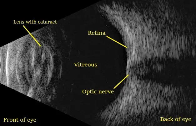
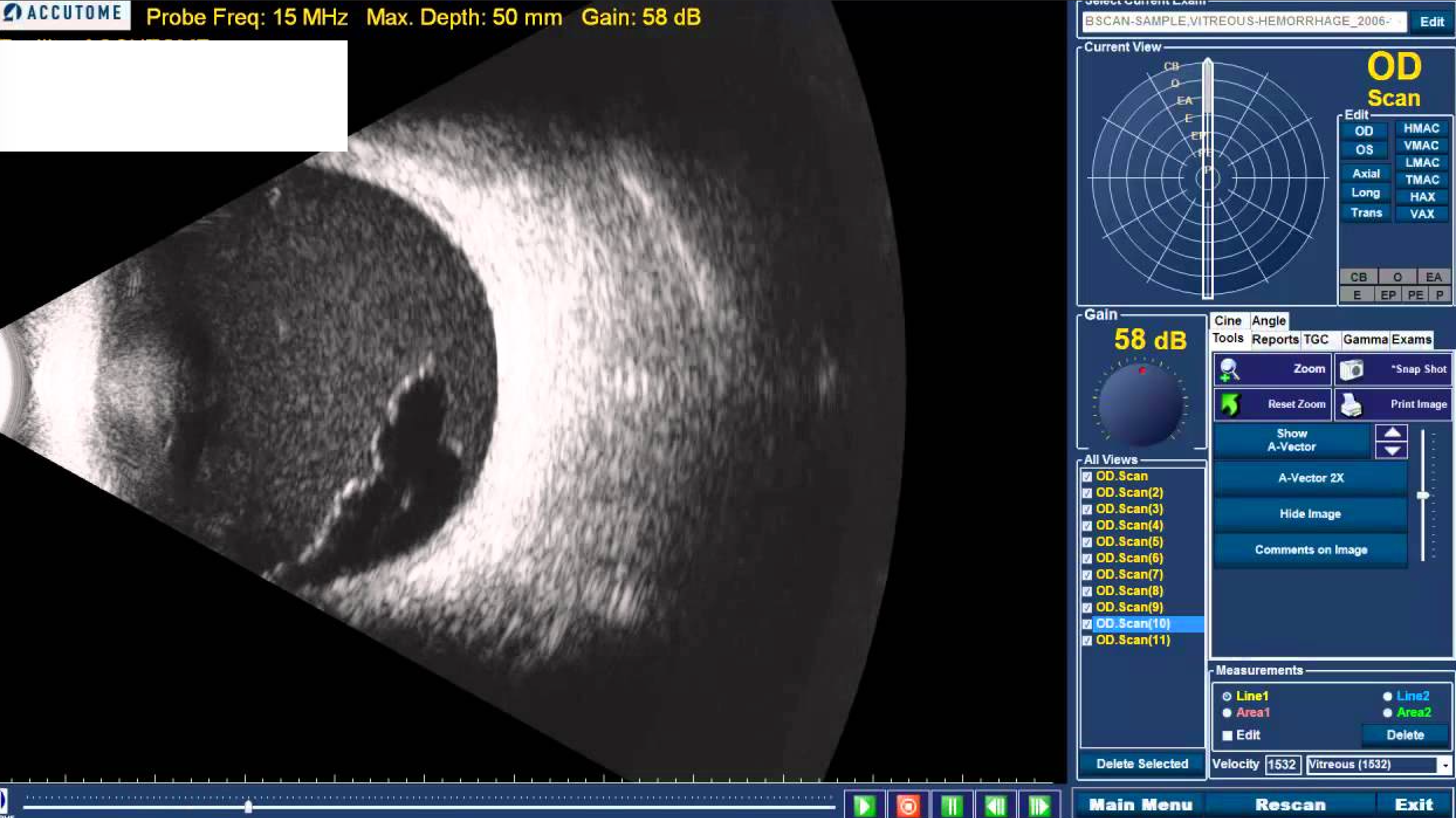
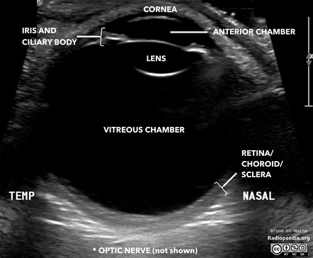
- Principle of ultrasonography: B-scan ultrasonography utilizes high- frequency sound waves (ultrasound) to create two-dimensional images of the internal structures of the eye. Sound waves are emitted from a transducer probe placed on the surface of the eye or eyelid, and they travel through the ocular tissues. When the sound waves encounter different tissue densities or interfaces within the eye, such as the boundaries between the vitreous, retina, and choroid, they are reflected back to the transducer probe.
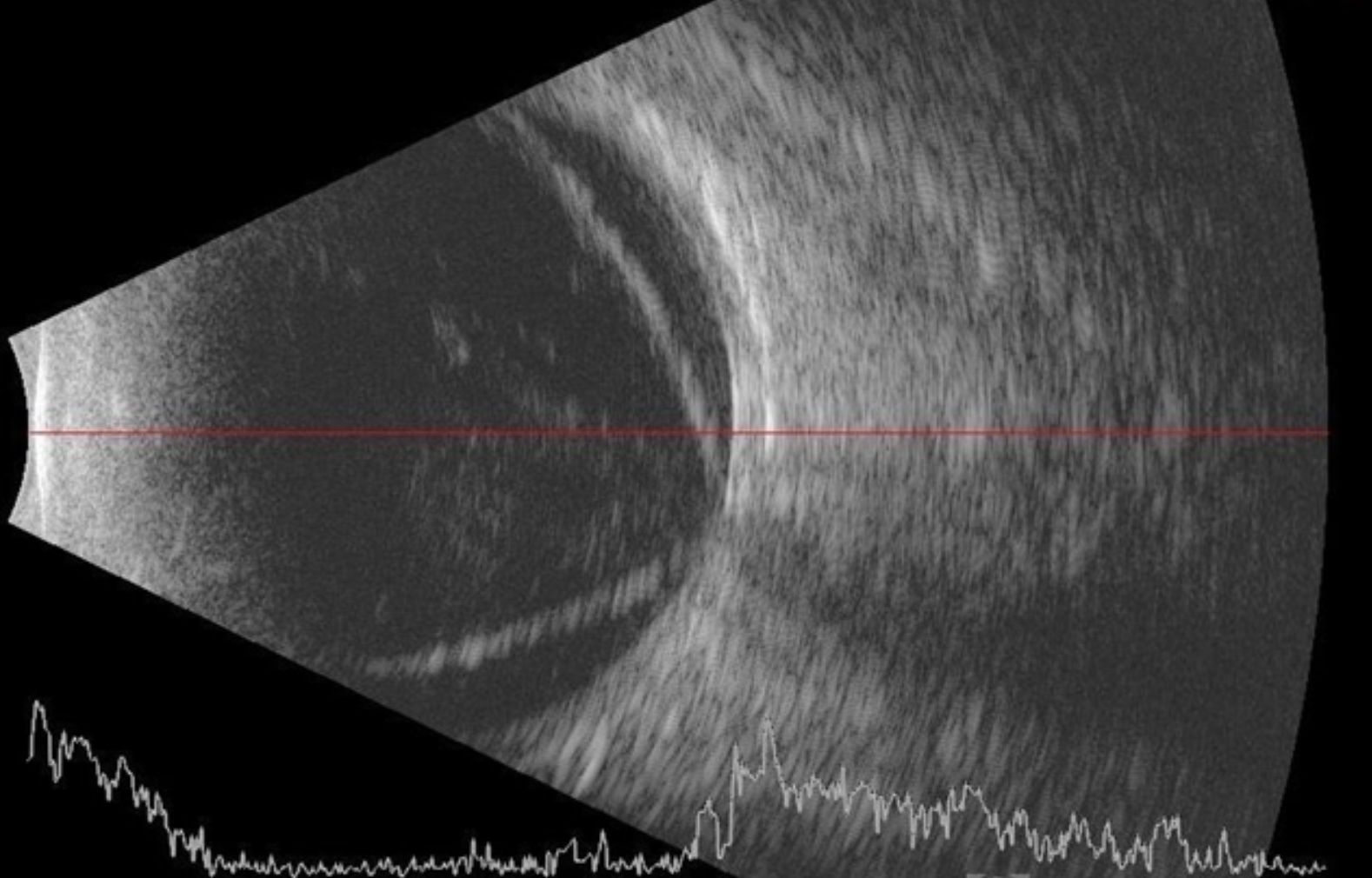
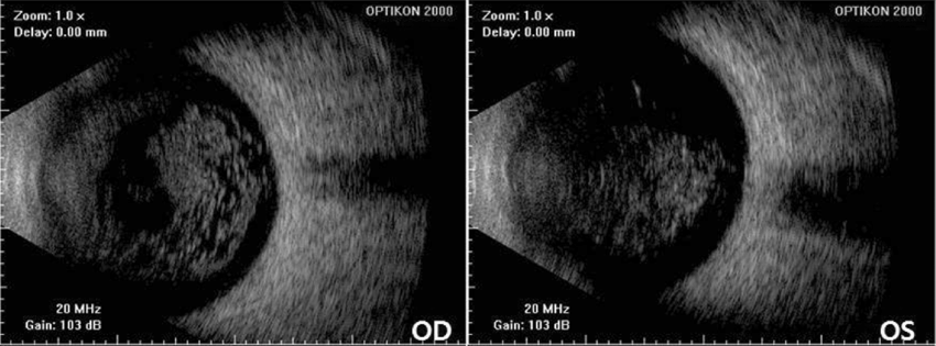
- Image formation: The reflected sound waves are then converted into electrical signals and processed by a computer to generate real-time images of the internal ocular structures. B-scan images are typically displayed as cross-sectional or longitudinal scans, allowing clinicians to visualize the thickness, shape, and position of various anatomical features.
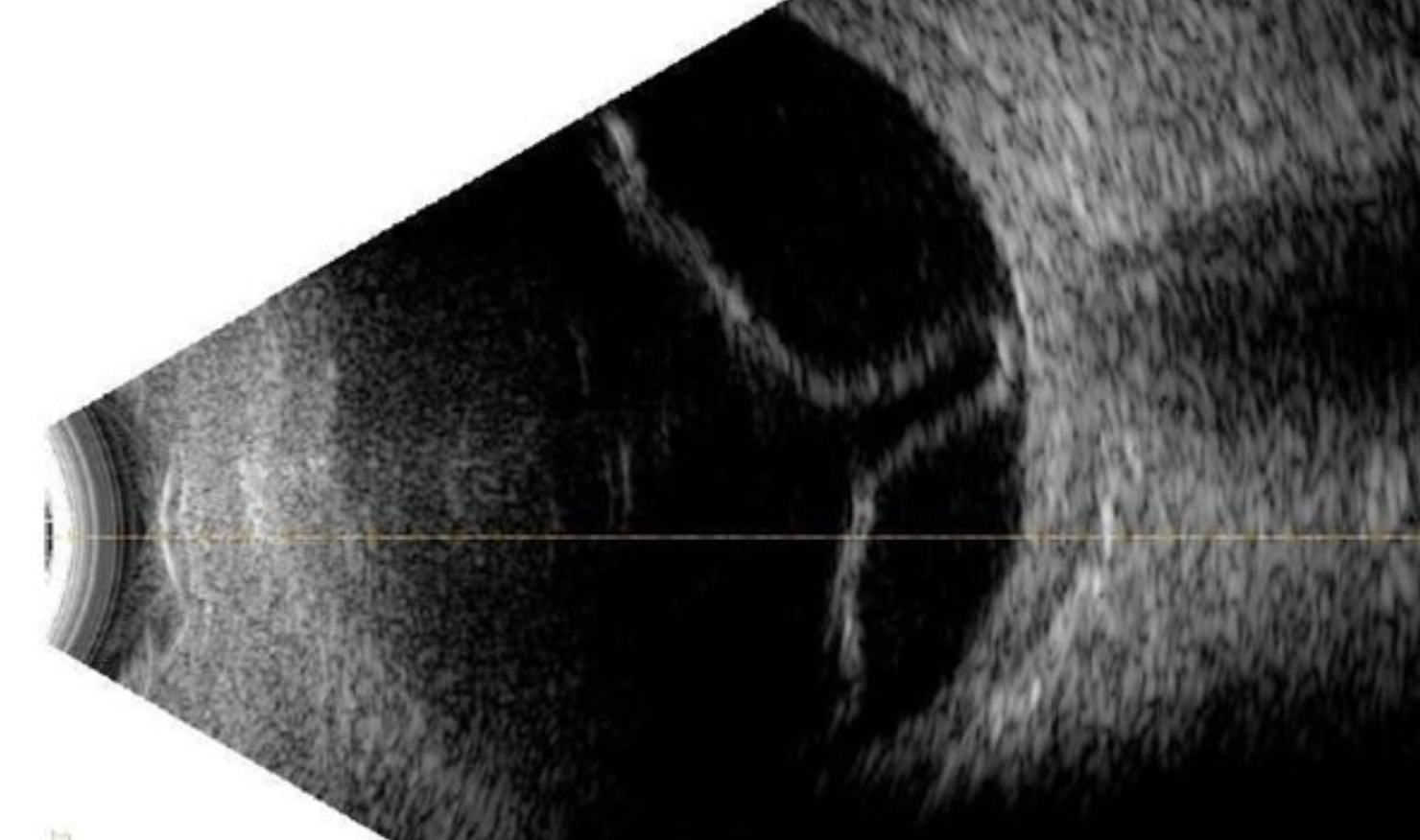
- Clinical applications: B-scan ultrasonography is used in a variety of clinical scenarios where direct visualization of the posterior segment of the eye is limited, such as in cases of opaque media (e.g., dense cataracts, corneal opacities) or in patients with poor pupillary dilation. It is particularly useful for evaluating conditions such as retinal detachments, vitreous hemorrhage, intraocular tumors (e.g., choroidal melanoma), foreign bodies, and ocular trauma. B-scan can also aid in assessing the position and integrity of intraocular implants (e.g., intraocular lenses) and guiding surgical interventions, such as vitrectomy or enucleation.
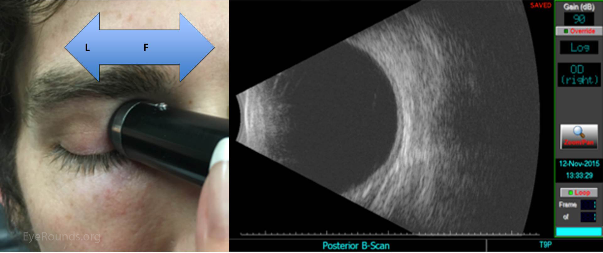
- Technique: During the B-scan examination, the patient is typically positioned in a reclined chair, and a coupling gel is applied to the closed eyelid or directly onto the transducer probe to ensure good acoustic contact and minimize air interface artifacts. The transducer probe is then gently placed on the eyelid or ocular surface, and the examiner maneuvers the probe to obtain multiple scans from different angles and orientations to thoroughly evaluate the ocular structures.
- Interpretation: B-scan images are interpreted by retina specialists who assess the morphology, location, and characteristics of any abnormalities or pathologies observed. B-scan findings are often correlated with clinical examination findings and other imaging modalities, such as optical coherence tomography (OCT) or fundus photography, to provide a comprehensive assessment of the ocular condition.