Optical Coherence Tomography
What is Optical Coherence Tomography?
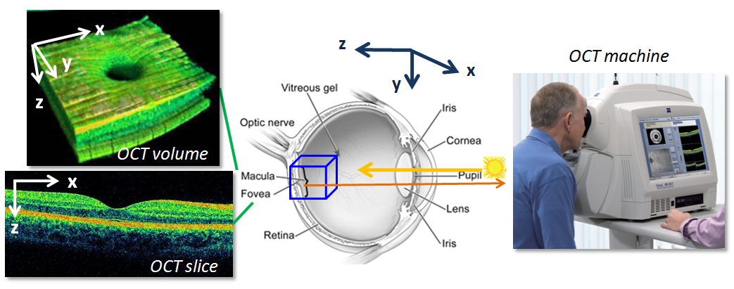
Optical Coherence Tomography (OCT) is a non-invasive imaging technique used to visualize and analyze the structures within the eye, particularly the retina. It provides high-resolution, cross-sectional images of these tissues, allowing retina specialists to assess their thickness, integrity, and morphology with exceptional detail.
Fundamentals of Optical Coherence Tomography
Here’s how OCT works and how it helps in clinical practice:
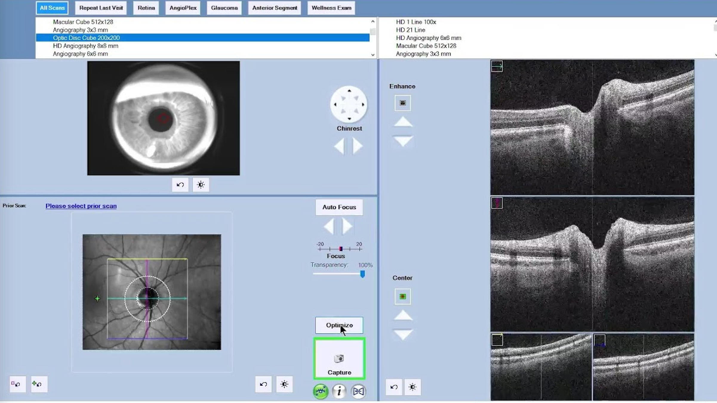
- Principle of OCT: OCT works on the principle of low-coherence interferometry. It utilizes a beam of light to scan the tissues of the eye, measuring the echo time delay and magnitude of light reflected from various structures. By analyzing the pattern of reflected light, OCT creates detailed, real-time images of the internal layers of the retina and other ocular structures.
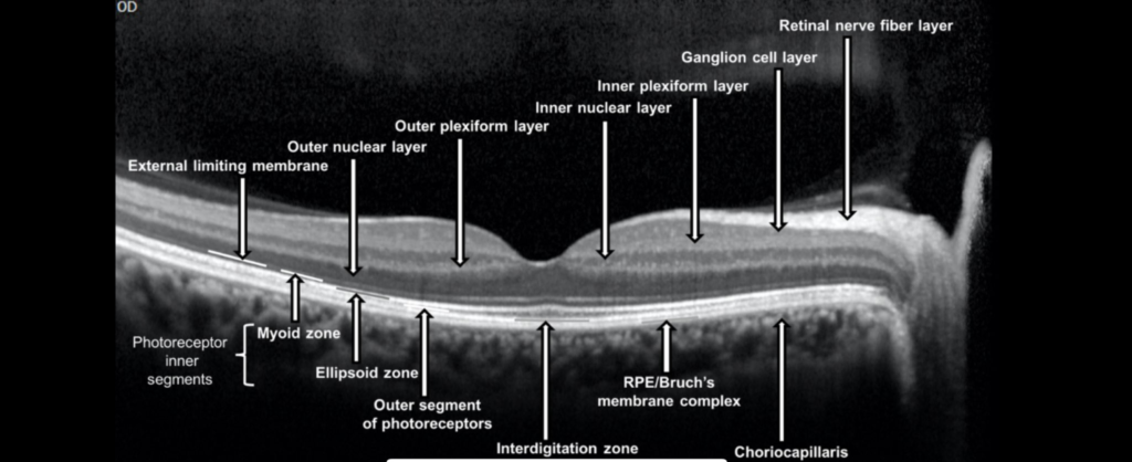
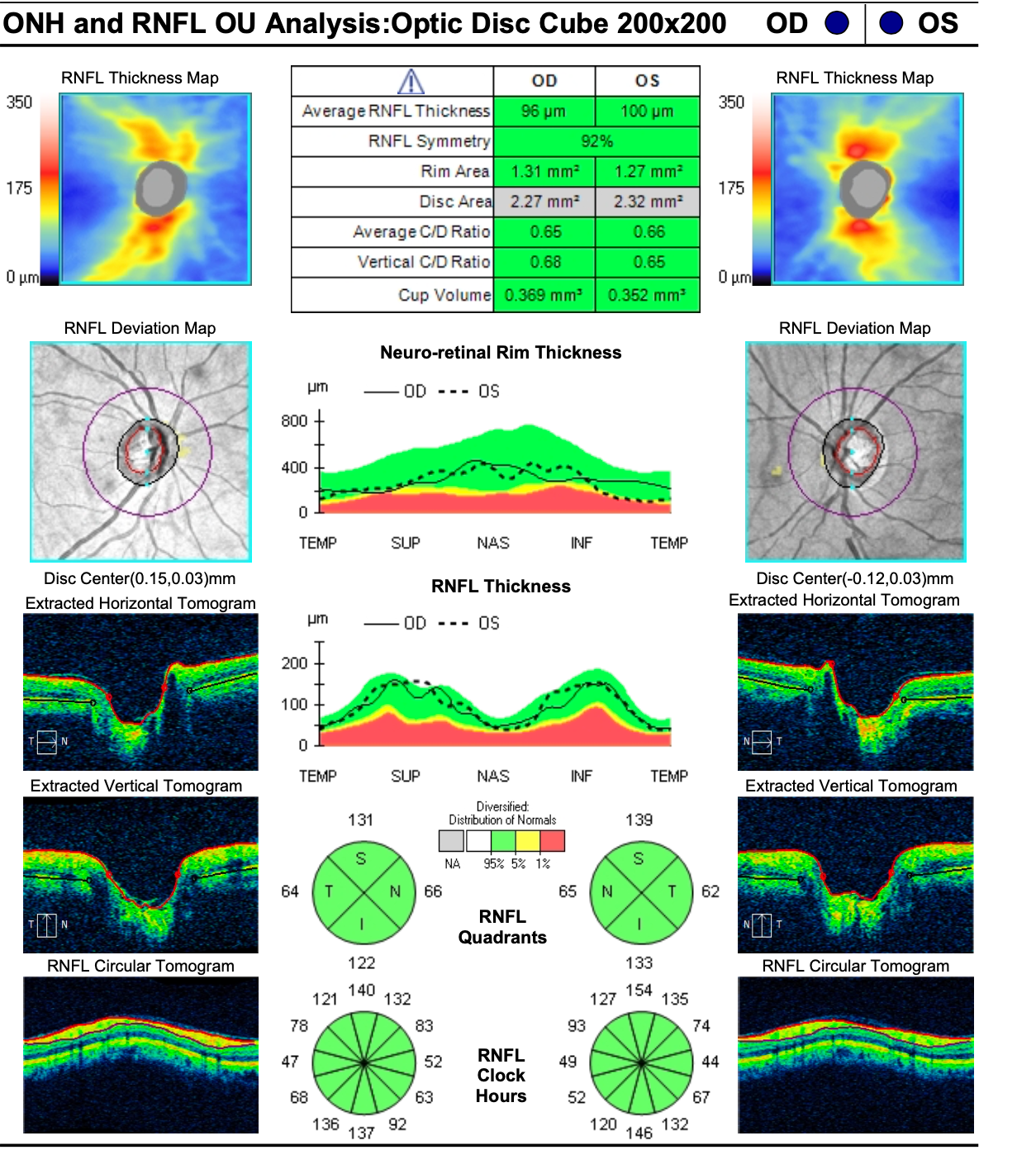
- Imaging capabilities: OCT can produce detailed, cross-sectional images of the retina, including its individual layers such as the retinal nerve fiber layer, ganglion cell layer, inner plexiform layer, outer plexiform layer, and photoreceptor layer. It can also visualize the choroid, the layer of blood vessels beneath the retina.
- Diagnostic applications: OCT is invaluable in the diagnosis and management of various retinal disorders. It helps retina specialists detect and monitor conditions such as age-related macular degeneration (AMD), diabetic retinopathy, macular edema, retinal vein occlusions, macular holes, and epiretinal membranes. By providing detailed information about the thickness, integrity, and morphology of retinal layers, OCT aids in the early detection of pathology, assessment of disease progression, and evaluation of treatment response.
- Treatment planning: In addition to diagnosis, OCT plays a crucial role in treatment planning for retinal and optic nerve disorders. It helps retina specialists identify the precise location and extent of pathology, guiding decisions regarding the need for interventions such as intravitreal injections, laser therapy, vitrectomy surgery, or glaucoma surgery. OCT measurements, such as central macular thickness and retinal nerve fiber layer thickness, can also be used to monitor the effectiveness of treatment over time.
- Research and education: OCT is widely used in clinical research and academic settings to investigate the pathophysiology of ocular diseases, evaluate the efficacy of new therapies, and advance our understanding of retinal and optic nerve anatomy and function. It is also an invaluable tool for teaching and training future generations of retina specialists, allowing them to visualize and interpret ocular pathology in a dynamic and interactive manner.
Overall, OCT is a versatile and indispensable imaging modality that has
revolutionized the field of ophthalmology, enabling precise diagnosis, personalized treatment, and enhanced patient care for a wide range of retinal and optic nerve disorders.