Retinal Detachment
What is Retinal Detachment?
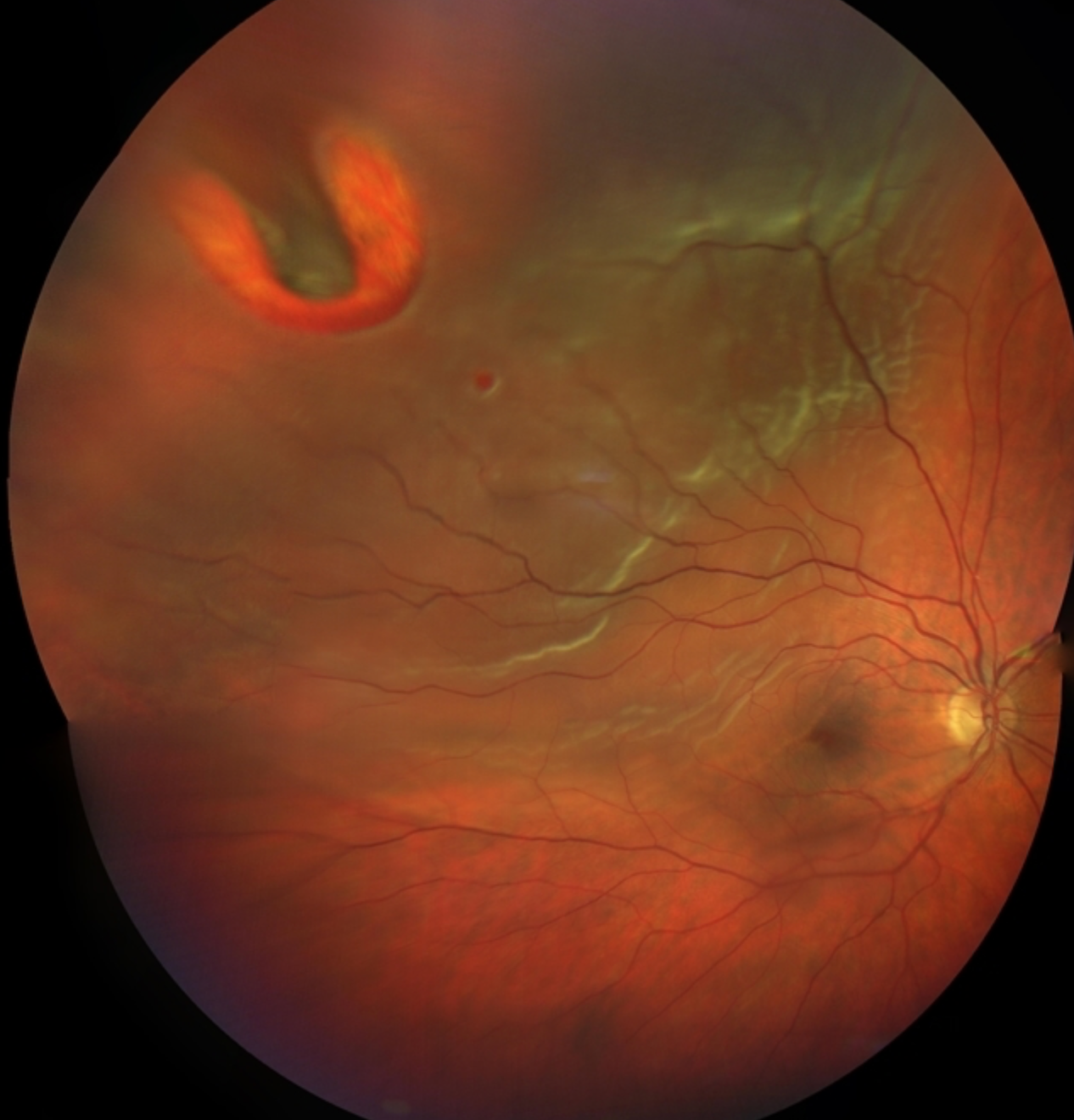
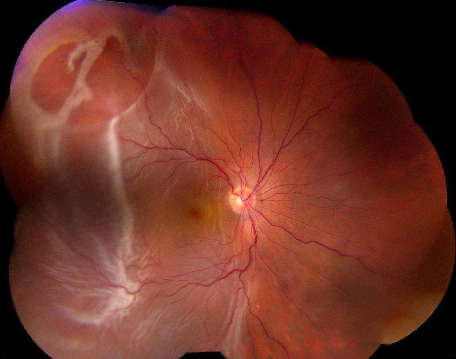
A retinal detachment is a serious eye condition where the retina, the light-sensitive tissue lining the back of the eye, pulls away from its normal position. This separation disrupts the blood supply to the retina, leading to vision loss if not promptly treated.
There are three main types of retinal detachment:
- Rhegmatogenous retinal detachment: This is the most common type and occurs when a tear or hole develops in the retina, allowing fluid from the vitreous gel to seep through and accumulate behind the retina, causing it to detach.
- Tractional retinal detachment: This type occurs when scar tissue on the retina contracts and pulls the retina away from the underlying tissue. Tractional retinal detachments are often associated with conditions such as diabetic retinopathy or proliferative vitreoretinopathy.
- Exudative retinal detachment: In this type, fluid accumulates underneath the retina without a tear or hole being present. Exudative retinal detachments are often caused by conditions such as age-related macular degeneration, inflammatory disorders, or tumors.
Symptoms of retinal detachment may include:
- Sudden onset of floaters (spots or cobwebs) in your field of vision
- Flashes of light in the affected eye, especially when moving your eyes
- A curtain-like shadow or veil across your field of vision
- Blurred vision or loss of vision in one eye
Treatments for Retinal Detachment
Treatment for retinal detachment typically involves surgical intervention to reattach the retina and prevent further vision loss. The choice of treatment depends on factors such as the type and severity of the detachment, as well as the patient’s overall eye health. Common surgical procedures for retinal detachment include:
- Vitrectomy: Vitrectomy is a surgical procedure where the vitreous gel inside the eye is removed and replaced with a clear solution. This allows the surgeon to directly access the retina and repair tears, remove scar tissue, or drain fluid from underneath the retina.
- Pneumatic retinopexy: In this procedure, a gas bubble is injected into the vitreous cavity to push the retina back into place and seal retinal tears. The patient then assumes a specific head position to keep the gas bubble in contact with the tear while it heals.
- Scleral buckle surgery: During this procedure, a silicone band or sponge is placed around the outside of the eye to gently push the wall of the eye inward, relieving traction on the retina. This helps close retinal tears and prevent fluid from seeping underneath the retina.
In some cases, a combination of these surgical techniques may be used to achieve the best outcome. It’s important to seek prompt medical attention if you experience symptoms of retinal detachment, as early detection and treatment can help prevent permanent vision loss.