Plaquenil (Hydroxychloroquine) Screening
What is Plaquenil (Hydroxychloroquine) Screening?
Plaquenil (hydroxychloroquine) is a medication used primarily to treat malaria, rheumatoid arthritis, and lupus. It works by reducing inflammation in the body and modulating the immune system. It can cause retinal toxicity primarily due to its accumulation in the retinal pigment epithelium (RPE) and photoreceptor cells (rods and cones). The exact mechanisms of retinal toxicity are not completely understood, but several factors are believed to contribute:
- Drug Accumulation: Hydroxychloroquine binds to melanin in the RPE, leading to prolonged retention and accumulation in retinal tissues.
- Phototoxicity: Hydroxychloroquine can absorb light and generate reactive oxygen species, leading to oxidative damage to retinal cells.
- Lysosomal Dysfunction: The drug can interfere with lysosomal function in the RPE, disrupting cellular homeostasis and leading to cell death.
- Direct Toxicity: Hydroxychloroquine may directly affect photoreceptor cells, causing apoptosis or programmed cell death.
Screening for Retinal Toxicity
Early detection of hydroxychloroquine-induced retinal toxicity is crucial to prevent irreversible vision loss. Current screening guidelines involve a combination of baseline and regular follow-up examinations using several techniques:
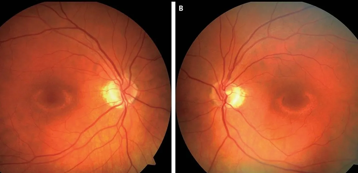
- Baseline Examination: Before starting hydroxychloroquine, a comprehensive eye examination is recommended, including:
- Visual acuity testing
- Dilated fundus examination
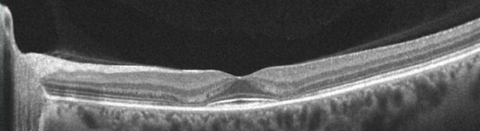
- Spectral-Domain Optical Coherence Tomography (SD-OCT): Provides high-resolution images of retinal layers to detect structural changes, such as loss of the photoreceptor inner segment/outer segment (IS/OS) junction.
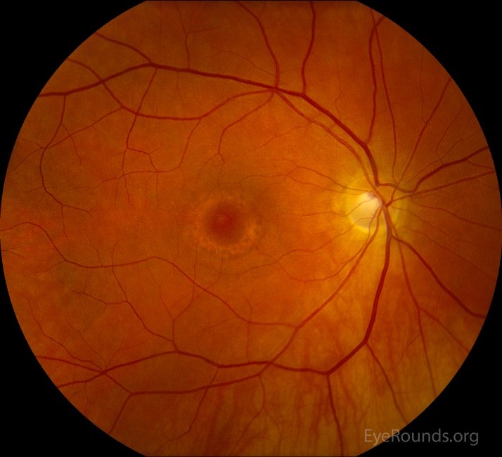
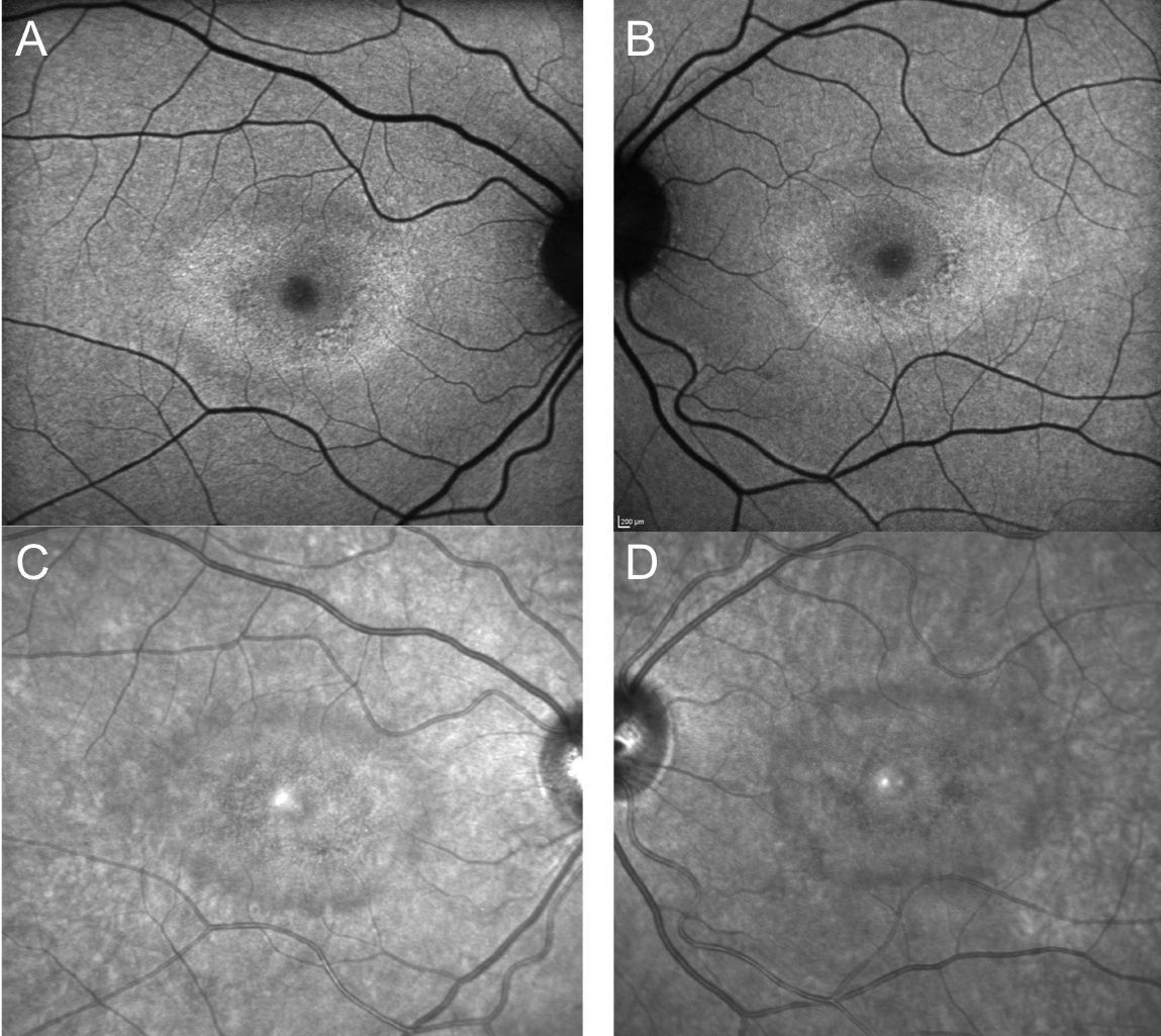
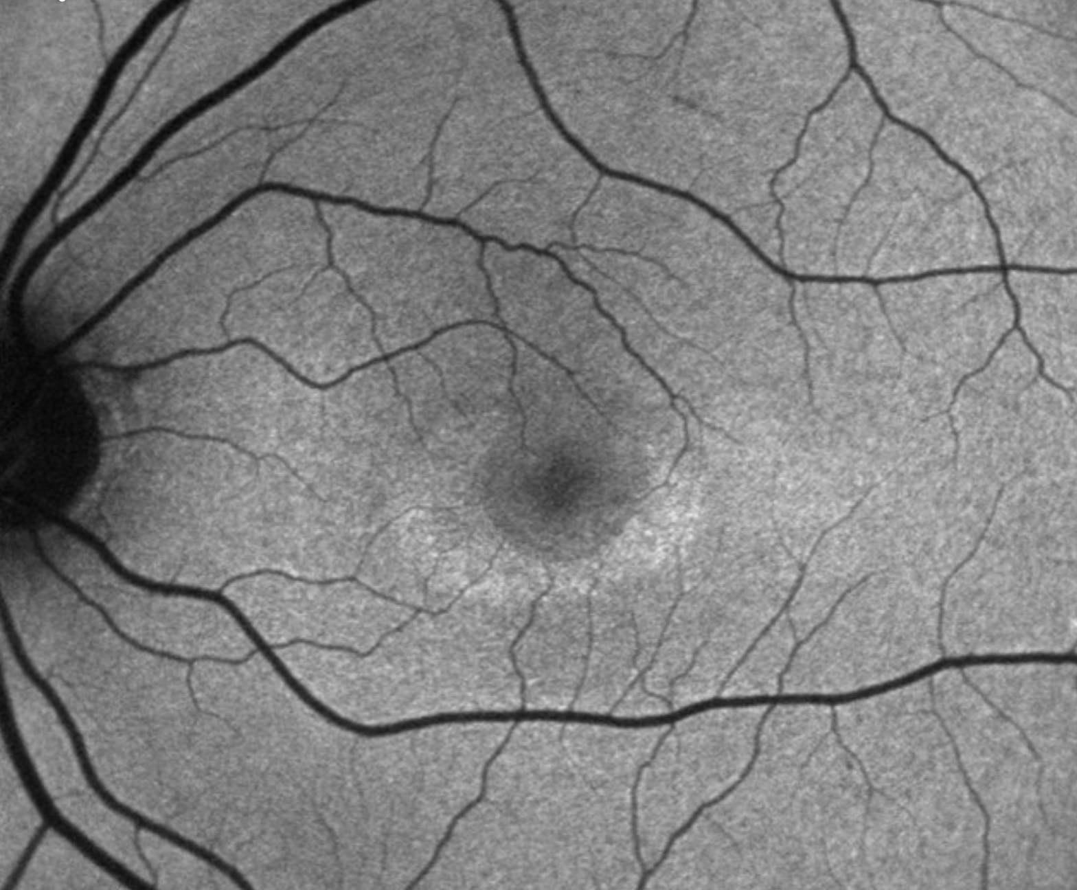
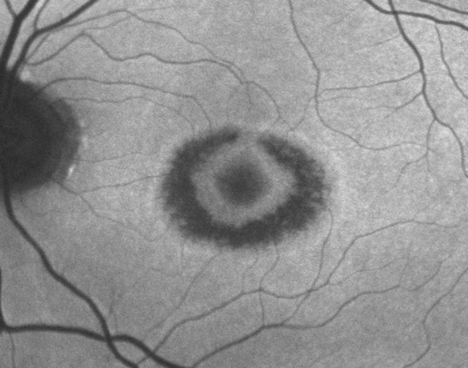
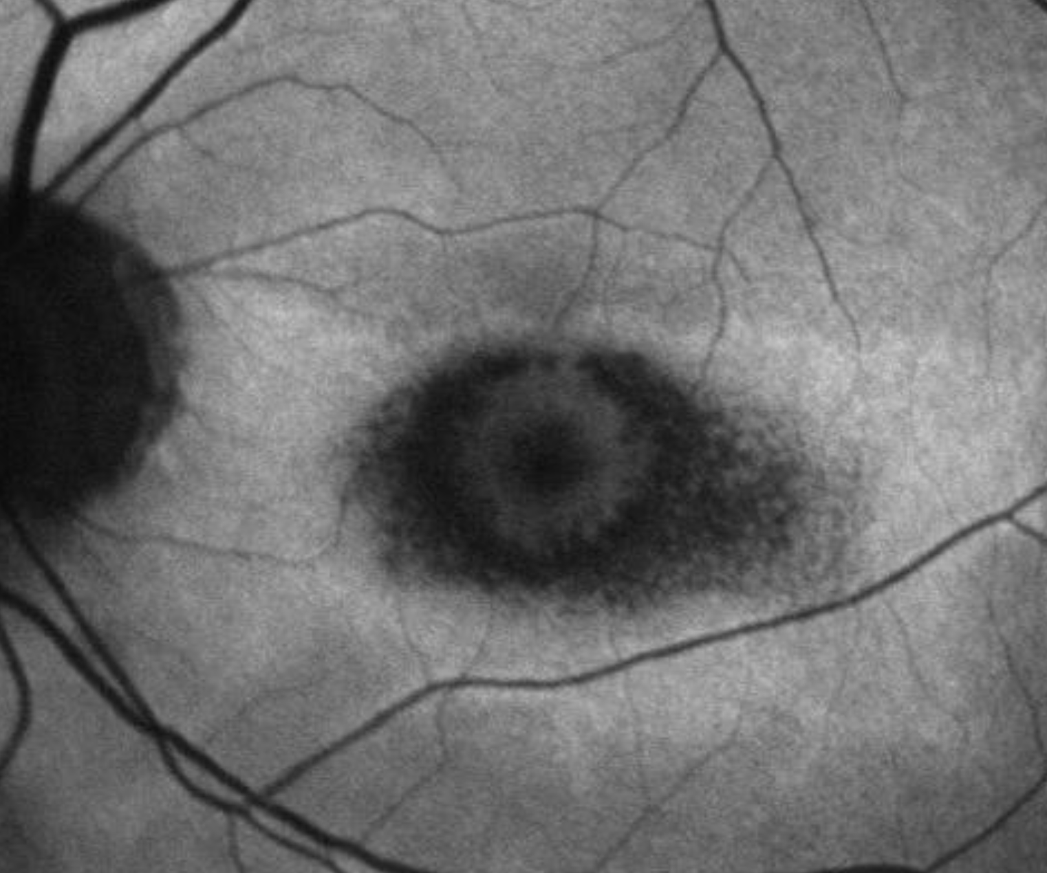
- Fundus Autofluorescence (FAF): Fundus Autofluorescence (FAF) is a non-invasive imaging technique used to assess the health of the retinal pigment epithelium (RPE) and photoreceptors in the retina. It is particularly useful in the detection and monitoring of hydroxychloroquine-induced retinal toxicity.
Detection of Hydroxychloroquine-Induced Retinal Toxicity
Early Detection of RPE Damage: Hydroxychloroquine toxicity primarily affects the RPE and photoreceptors. FAF can reveal early abnormalities in the RPE before they are visible on clinical examination or other imaging modalities. Areas of increased autofluorescence may indicate stressed or damaged RPE cells, while areas of decreased autofluorescence can indicate RPE atrophy. Patterns of Autofluorescence: Specific patterns of autofluorescence changes are associated with hydroxychloroquine toxicity: Pericentral Hyperfluorescence: Early signs of toxicity might show as a ring of increased autofluorescence around the macula, corresponding to areas of early RPE stress. Parafoveal Changes: More advanced stages may show a “bull’s eye” pattern, characterized by a ring of reduced autofluorescence around the fovea, indicating RPE and photoreceptor loss.
Risk Factors for Hydroxychloroquine-Induced Retinal Toxicity
Certain factors increase the risk of hydroxychloroquine-induced retinal toxicity:
- High Dosage: Doses exceeding 5 mg/kg/day of actual body weight.
- Duration of Use: Use of the drug for more than five years.
- Pre-existing Retinal Disease: Pre-existing macular disease or other retinal conditions.
- Kidney or Liver Dysfunction: Reduced clearance of the drug leading to higher retinal concentrations.
- Concurrent Use of Tamoxifen: Tamoxifen can potentiate retinal toxicity.